immunofluorescence staining - immunofluorescent photomicrograph stock pictures, royalty-free photos & imagesImmunofluorescence stainingMagnification, Fluorescent, Endothelial
immunofluorescence microscopy - immunofluorescent photomicrograph stock pictures, royalty-free photos & imagesImmunofluorescence Microscopy
immunoflourescence of a smooth muscle cell - immunofluorescent photomicrograph stock pictures, royalty-free photos & imagesImmunoflourescence of a smooth muscle cellFluorescent, Magnification, Histology
multiphoton fluorescence image of hela cells. - immunofluorescent photomicrograph stock pictures, royalty-free photos & imagesMultiphoton fluorescence image of HeLa cells.HeLa Cells, DNA, Fluorescent
female researcher using inverted microscope to view stem cells displayed showing fluorescent labeled cells - immunofluorescent photomicrograph stock pictures, royalty-free photos & imagesFemale researcher using inverted microscope to view stem cells displayed showing fluorescent labeled cellsMicroscope, Fluorescent, Scientist
slide demonstrating breast tissue with ductal carcinoma. histopathology on the microscope. immunofluorescent photomicrograph, organs samples, histological examination, - immunofluorescent photomicrograph stock pictures, royalty-free photos & imagesSlide demonstrating breast tissue with ductal carcinoma. Histopathology on the microscope. Immunofluorescent photomicrograph, Organs samples, Histological examination,Bone Marrow Tissue, Microscope, Abstract
Using phase contrast imaging and magnified 1125X, this photomicrograph depicts a cyst of the protozoan Acanthamoeba polyphaga. CDC/Dr George Healy...Acanthamoeba polyphaga
Photo Essay In Curie Institute, At Nikon Imaging Centre, Paris, France. In Vivo Photonic Imagery Platform For Biomedical Research To Observe Alive...Rab2
Photo Essay In Curie Institute, At Nikon Imaging Centre, Paris, France. In Vivo Photonic Imagery Platform For Biomedical Research To Observe Alive...Confocal Microscope
biological mouse tissue, from the bone area, education anatomy and histological sample simple columnar epithelium tissue under the microscope. stock photo - immunofluorescent photomicrograph stock pictures, royalty-free photos & imagesBiological mouse tissue, from the bone area, education anatomy and Histological sample Simple columnar epithelium Tissue under the microscope. stock photoSkin, Analysing, Microscope
incubation of cells with cytostatics, actin filaments(nanotechnology) and morphological changes following chemotherapy using confocal microscopy - immunofluorescent photomicrograph stock pictures, royalty-free photos & imagesIncubation of cells with cytostatics, actin filaments(nanotechnology) and morphological changes following chemotherapy using confocal microscopyMitochondrion, Fluorescent, Research
mesenchymal stem cells labeled with fluorescent molecules. - immunofluorescent photomicrograph stock pictures, royalty-free photos & imagesMesenchymal stem cells labeled with fluorescent molecules.Cancer Cell, Abstract, Contrasts
sampling strategy of the stem tissues. immunofluorescent photomicrograph, organs samples, histological examination, histopathology on the microscope - immunofluorescent photomicrograph stock pictures, royalty-free photos & imagesSampling strategy of the stem tissues. Immunofluorescent photomicrograph, Organs samples, Histological examination, histopathology on the microscopeMicroscope, Plant, Magnification
indirect immunofluorescence test, illustration - immunofluorescent photomicrograph stock illustrationsIndirect immunofluorescence test, illustrationConcepts
simple cuboidal epithelium in a kidney tubule, 100x - immunofluorescent photomicrograph stock pictures, royalty-free photos & imagesSIMPLE CUBOIDAL EPITHELIUM in a KIDNEY TUBULE, 100XTubules, Epithelium
microscopic photo of a professionally prepared slide demonstrating breast tissue - immunofluorescent photomicrograph stock pictures, royalty-free photos & imagesMicroscopic photo of a professionally prepared slide demonstrating breast tissueDNA, Microscope, People
aspergillus (mold). conidia & conidiophores, 250x at 35mm. the circular structures are conidia (spores) that pinch off. asexual reproduction. aspergillus is a deuteromycet. - immunofluorescent photomicrograph stock pictures, royalty-free photos & imagesAspergillus (mold). Conidia & conidiophores, 250X at 35mm. The circular structures are conidia (spores) that pinch off. Asexual Reproduction. Aspergillus is a deuteromycet.Magnification, Fungus, Photography
motor neuron (multipolar) with cell body and many processes (mostly dendrites), spinal cord (magnification x100). this multipolar motor neuron comes from the anterior (ventral) horn of the spinal cord grey matter. the stain is methylene blue and phloxine. - immunofluorescent photomicrograph stock pictures, royalty-free photos & imagesMotor Neuron (Multipolar) with Cell Body and many Processes (mostly Dendrites), Spinal Cord (Magnification x100). This multipolar motor neuron comes from the anterior (ventral) horn of the spinal cord grey matter. The stain is methylene blue and phloxine.Microscope, Human Cell, Scientific Micrograph
rodadas, granada 2017 - immunofluorescent photomicrograph stock pictures, royalty-free photos & imagesRodadas, Granada 2017
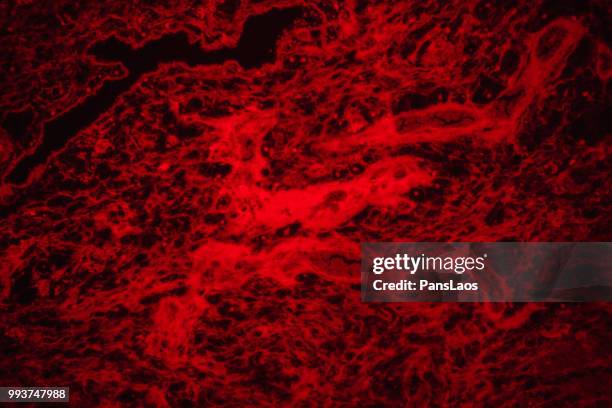
medical hemangioma tumor - immunofluorescent photomicrograph stock pictures, royalty-free photos & imagesmedical Hemangioma tumorMicroscope
medical hemangioma tumor - immunofluorescent photomicrograph stock pictures, royalty-free photos & imagesmedical Hemangioma tumorPeople, Melanocyte, Melanoma
medical hemangioma tumor - immunofluorescent photomicrograph stock pictures, royalty-free photos & imagesmedical Hemangioma tumorMicroscope, Haemorrhage, Blood
medical hemangioma tumor - immunofluorescent photomicrograph stock pictures, royalty-free photos & imagesmedical Hemangioma tumor
medical hemangioma tumor - immunofluorescent photomicrograph stock pictures, royalty-free photos & imagesmedical Hemangioma tumor
medical hemangioma tumor - immunofluorescent photomicrograph stock pictures, royalty-free photos & imagesmedical Hemangioma tumor
medical hemangioma tumor - immunofluorescent photomicrograph stock pictures, royalty-free photos & imagesmedical Hemangioma tumorDermis
medical hemangioma tumor - immunofluorescent photomicrograph stock pictures, royalty-free photos & imagesmedical Hemangioma tumor
medical hemangioma tumor - immunofluorescent photomicrograph stock pictures, royalty-free photos & imagesmedical Hemangioma tumorCancer Cell, Blood
medical hemangioma tumor - immunofluorescent photomicrograph stock pictures, royalty-free photos & imagesmedical Hemangioma tumorPeople, Tissue - Anatomy
medical hemangioma tumor - immunofluorescent photomicrograph stock pictures, royalty-free photos & imagesmedical Hemangioma tumor
medical hemangioma tumor - immunofluorescent photomicrograph stock pictures, royalty-free photos & imagesmedical Hemangioma tumor
medical hemangioma tumor - immunofluorescent photomicrograph stock pictures, royalty-free photos & imagesmedical Hemangioma tumorCancer Cell, Microscope, People
medical hemangioma tumor - immunofluorescent photomicrograph stock pictures, royalty-free photos & imagesmedical Hemangioma tumor
medical hemangioma tumor - immunofluorescent photomicrograph stock pictures, royalty-free photos & imagesmedical Hemangioma tumorBlood, Illness
fireworks on the coast - immunofluorescent photomicrograph stock pictures, royalty-free photos & imagesFireworks on the CoastCytoskeleton
epithelium--transitional epithelium lining the inside of the urinary bladder, 100x - immunofluorescent photomicrograph stock pictures, royalty-free photos & imagesEPITHELIUM--TRANSITIONAL EPITHELIUM lining the inside of the Urinary Bladder, 100X
zigzag old rubber wheel profile - immunofluorescent photomicrograph stock pictures, royalty-free photos & imageszigzag old rubber wheel profile
bon nadal, felices fiestas! - immunofluorescent photomicrograph stock pictures, royalty-free photos & imagesBon Nadal, Felices Fiestas!
late winter contrast - immunofluorescent photomicrograph stock pictures, royalty-free photos & imagesLate Winter Contrast
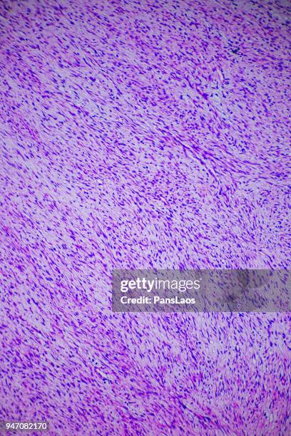
fibroma tumor cells medical micrograph of ill human - immunofluorescent photomicrograph stock pictures, royalty-free photos & imagesFibroma tumor cells medical micrograph of ill human
fibroma tumor cells medical micrograph of ill human - immunofluorescent photomicrograph stock pictures, royalty-free photos & imagesFibroma tumor cells medical micrograph of ill human
fibroma tumor cells medical micrograph of ill human - immunofluorescent photomicrograph stock pictures, royalty-free photos & imagesFibroma tumor cells medical micrograph of ill human
fibroma tumor cells medical micrograph of ill human - immunofluorescent photomicrograph stock pictures, royalty-free photos & imagesFibroma tumor cells medical micrograph of ill human
fibroma tumor cells medical micrograph of ill human - immunofluorescent photomicrograph stock pictures, royalty-free photos & imagesFibroma tumor cells medical micrograph of ill humanGynaecologist, Hysterectomy, Cancer Cell
fibroma tumor cells medical micrograph of ill human - immunofluorescent photomicrograph stock pictures, royalty-free photos & imagesFibroma tumor cells medical micrograph of ill human
White blood cells , a stack of platelets, and red blood cells revealed in the photomicrograph of a thick film Giemsa-stained blood smear sample,...White And Red Blood CellsPurple
papillae on the human tongue, 5x - immunofluorescent photomicrograph stock pictures, royalty-free photos & imagesPAPILLAE on the human TONGUE, 5X
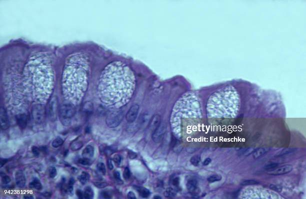
goblet cells and simple columnar epithelium in the large intestine 250x - immunofluorescent photomicrograph stock pictures, royalty-free photos & imagesGOBLET CELLS and Simple Columnar Epithelium in the Large Intestine 250XGoblet Cell
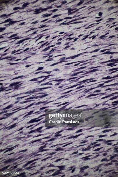
smooth muscle teased apart to show the anatomy of individual cells or fibers, 100x - immunofluorescent photomicrograph stock pictures, royalty-free photos & imagesSMOOTH MUSCLE teased apart to show the anatomy of individual cells or fibers, 100X
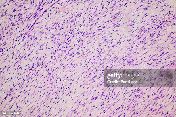
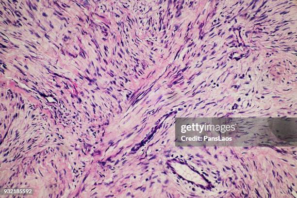
fibroma tumour of human micrograph - immunofluorescent photomicrograph stock pictures, royalty-free photos & imagesFibroma tumour of human micrographPeople, Magnification
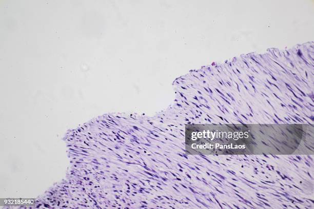
medical fibroma tumour of human - immunofluorescent photomicrograph stock pictures, royalty-free photos & imagesmedical fibroma tumour of humanHuman Cell, Tissue - Anatomy, Magnification
medical fibroma tumour of human - immunofluorescent photomicrograph stock pictures, royalty-free photos & imagesmedical fibroma tumour of human
fibroma tumour of human micrograph - immunofluorescent photomicrograph stock pictures, royalty-free photos & imagesFibroma tumour of human micrograph
fibroma tumour of human - immunofluorescent photomicrograph stock pictures, royalty-free photos & imagesfibroma tumour of human
medical science fluorescence micrograph of human fibroma tumour - immunofluorescent photomicrograph stock pictures, royalty-free photos & imagesmedical science fluorescence micrograph of human fibroma tumourUterus
fibroma tumour of human micrograph - immunofluorescent photomicrograph stock pictures, royalty-free photos & imagesFibroma tumour of human micrographPeople, Magnification
fibroma tumour of human - immunofluorescent photomicrograph stock pictures, royalty-free photos & imagesfibroma tumour of humanLeiomyoma
fibroma tumour of human micrograph - immunofluorescent photomicrograph stock pictures, royalty-free photos & imagesFibroma tumour of human micrographDermis