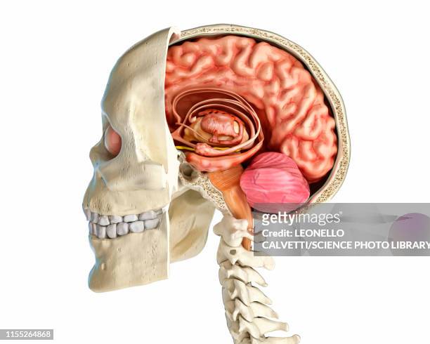
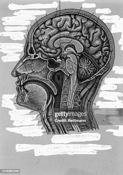
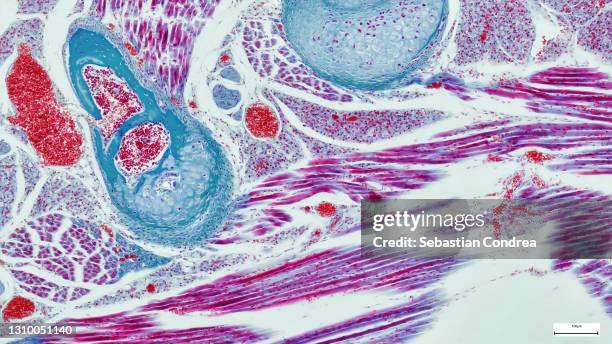
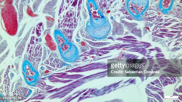
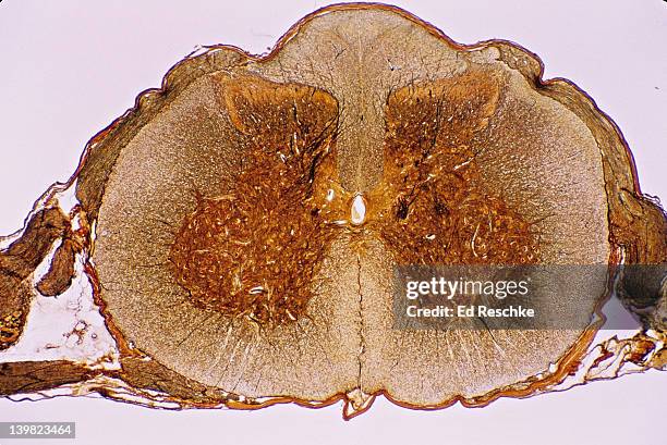
spinal cord cross section, 5x shows: gray matter (golden butterfly), white matter, central canal, dorsal & ventral root, dorsal root ganglion, meninges, dorsal horn, ventral (anterior) horn, & anterior horn cells (motor neuron cell bodies) - brain cross section stock pictures, royalty-free photos & images
hippocampal sulcus remnant cysts seen on t2 axial mri (magnetic resonance imaging) - brain cross section stock pictures, royalty-free photos & images
stem cross section. buttercup (ranunculus), herbaceous dicot, 8x. shows: vascular bundles arranged in a ring (typical of dicots), xylem, phloem, epidermis, cortex, and pith. - brain cross section stock pictures, royalty-free photos & images
digital illustration of human brain showing lobes and cross section - brain cross section stock illustrations
human anatomy, cross section of head showing brain, cerebrum, cerebellum, pons varolii, spinal cord - brain cross section stock illustrations
micrograph of rat brain. science cross section. - brain cross section stock pictures, royalty-free photos & images
micrograph of rat brain. science cross section. immunofluorescent photomicrograph, organs samples, histological examination, histopathology on the microscope - brain cross section stock pictures, royalty-free photos & images
stem cross section. sunflower (helianthus), herbaceous dicot, 10x. shows: vascular bundles arranged in a ring, zylem, phloem, pith, cortex, and epidermis. - brain cross section stock pictures, royalty-free photos & images
Cerebral atrophy, anterior temporal and parietal frontal Ponto cerebellar, radial cross-section MRI cranial scan.
lateral cross section through the skull showing the brain with intermediate layers (meninges) highlighted. - brain cross section stock illustrations
Anatomical Model Of The Human Brain Or Encephalon Cross Section. The Encephalon Is Composed Of The Diencephalon, Covered By The Cerebrum Flesh...
stem cross section. alfalfa (medicago), herbaceous dicot, 25x. vascular bundles are arranged in a ring. shows: vascular bundles, xylem, phloem, epidermis, cortex, pith & collenchyma in the corners. - brain cross section stock pictures, royalty-free photos & images
Slices of a human brain donated from a 56-year-old male who had "active MS," Samples of human brains at the Department of Neurosurgery at the...
blue x-ray image of human brain anatomy, 3-d sagittal section (side view, cross section) of human brain and its parts - brain cross section stock pictures, royalty-free photos & images
spinal cord. cross section, 5x shows: gray matter (inner pink, butterfly-shaped area), white matter (outer blue area), central canal, meninges, dorsal horn, lateral horn, ventral (anterior) horn, and anterior horn cells (motor neuron cell bodies). - brain cross section stock pictures, royalty-free photos & images
cross section of the cerebellum and nerve human under the microscope for education in lab - brain cross section stock pictures, royalty-free photos & images
cross section illustration of human brain showing limbic system and primitive forebrain - brain cross section stock illustrations
Cerebral atrophy. Parietal cortico-sub-cortical atrophies , seen on a radial cross-section brain MRI scan.
Cerebral atrophy, anterior temporal and parietal frontal Ponto cerebellar, radial cross-section MRI cranial scan.
Cerebrovascular accident caused by thrombosis of an artery in the left hemisphere. Saggital plane cross-section brain scan.
Cerebral atrophy. Parietal cortico-sub-cortical atrophies , seen on a sagittal plane cross-section brain CT scan.
cross section - xylem is a type of tissue in vascular plants that transports water and some nutrients. scientific research. plant tissue structureimmunofluorescent photomicrograph, organs samples, histological examination, histopathology on the microscope - brain cross section stock pictures, royalty-free photos & images
Black and white print illustrating a cross-section of the lateral ventricles of a human brain, with alphabetized figures indicating the sites of...
Cerebrovascular accident caused by thrombosis of an artery in the left hemisphere. Saggital plane cross-section brain scan.
x-section of buttercup mature root. ranunculus. shows stele, xylem, phloem, cortex, endodermis, starch grains & epidermis. dicot 10x - brain cross section stock pictures, royalty-free photos & images
microscopic image of kidney cross section - brain cross section stock pictures, royalty-free photos & images
Cerebral atrophy. Parietal cortico-sub-cortical atrophies , seen on a sagittal plane cross-section brain CT scan.
Cerebral atrophy - Posterior cortical atrophy (posterior cortical degeneration responsible for agnosia, apraxia, aphasia, Gerstmann syndrome,...
close-up of a cross section of a red cabbage - brain cross section stock pictures, royalty-free photos & images
Phrenological cross-section of a man's head, illustrating the idea that the brain processes thoughts in different locations according to their type.
stem cross section of brazilian waterweed (egeria densa) showing cortex, pith and vascular tissue. magnified 60x (ref. at 35 mm film frame) - brain cross section stock pictures, royalty-free photos & images
Cerebral atrophy, anterior temporal and parietal frontal Ponto cerebellar, radial cross-section MRI cranial scan.
Cerebral atrophy, anterior temporal and parietal frontal Ponto cerebellar, frontal cross-section MRI cranial scan.
Cerebral atrophy, anterior temporal and parietal frontal Ponto cerebellar, radial cross-section MRI cranial scan.
Cerebral atrophy, anterior temporal and parietal frontal Ponto cerebellar, frontal cross-section MRI cranial scan.